BMRB Entry 16882
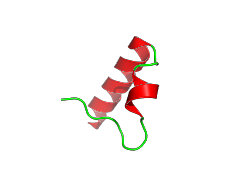
Click here to enlarge.
PDB ID:
Entry in NMR Restraints Grid
Validation report in NRG-CING
Chem Shift validation: AVS_full, LACS
BMRB Entry DOI: doi:10.13018/BMR16882
MolProbity Validation Chart
NMR-STAR file interactive viewer.
NMR-STAR v3 text file.
XML gzip file.
RDF gzip file.
All files associated with the entry
Citation: Bomar, Martha; Bienko, Sanjay; Dikic, Marzena; Walker, Ivan; Zhou, Graham. "Unconventional ubiquitin recognition by the ubiquitin-binding motif within the Y family DNA polymerases iota and Rev1." Mol. Cell 37, 408-417 (2010).
PubMed: 20159559
Assembly members:
Ubiquitin-Binding Motif, polymer, 108 residues, 3753.299 Da.
Natural source: Common Name: Human Taxonomy ID: 9606 Superkingdom: Eukaryota Kingdom: Metazoa Genus/species: Homo sapiens
Experimental source: Production method: recombinant technology Host organism: Escherichia coli Vector: pET30-GB1-fusion
Entity Sequences (FASTA):
Ubiquitin-Binding Motif: MQYKLILNGKTLKGETTTEA
VDAATAEKVFKQYANDNGVD
GEWTYDDATKTFTVTEGSDE
KITFPSDIDPQVFYELPEAV
QKELLAEWKRTGSDFHIGHK
LEHHHHHH
- assigned_chemical_shifts
| Data type | Count |
| 13C chemical shifts | 435 |
| 15N chemical shifts | 103 |
| 1H chemical shifts | 694 |
Additional metadata:
Assembly:
| Entity Assembly ID | Entity Name | Entity ID |
|---|---|---|
| 1 | UBM2 | 1 |
Entities:
Entity 1, UBM2 108 residues - 3753.299 Da.
Residues 1-56 represent the GB1-tag. Residues 57-58, 101-108 are cloning artifacts (including the His6-tag)
| 1 | MET | GLN | TYR | LYS | LEU | ILE | LEU | ASN | GLY | LYS | ||||
| 2 | THR | LEU | LYS | GLY | GLU | THR | THR | THR | GLU | ALA | ||||
| 3 | VAL | ASP | ALA | ALA | THR | ALA | GLU | LYS | VAL | PHE | ||||
| 4 | LYS | GLN | TYR | ALA | ASN | ASP | ASN | GLY | VAL | ASP | ||||
| 5 | GLY | GLU | TRP | THR | TYR | ASP | ASP | ALA | THR | LYS | ||||
| 6 | THR | PHE | THR | VAL | THR | GLU | GLY | SER | ASP | GLU | ||||
| 7 | LYS | ILE | THR | PHE | PRO | SER | ASP | ILE | ASP | PRO | ||||
| 8 | GLN | VAL | PHE | TYR | GLU | LEU | PRO | GLU | ALA | VAL | ||||
| 9 | GLN | LYS | GLU | LEU | LEU | ALA | GLU | TRP | LYS | ARG | ||||
| 10 | THR | GLY | SER | ASP | PHE | HIS | ILE | GLY | HIS | LYS | ||||
| 11 | LEU | GLU | HIS | HIS | HIS | HIS | HIS | HIS |
Samples:
sample_1: UBM2, [U-100% 13C; U-100% 15N], 1 – 4 mM; D2O 100%
sample_conditions_1: ionic strength: 0.1 M; pH: 7; pressure: 1 atm; temperature: 298 K
Experiments:
| Name | Sample | Sample state | Sample conditions |
|---|---|---|---|
| 3D HCCH-TOCSY | sample_1 | isotropic | sample_conditions_1 |
| 3D HNCA | sample_1 | isotropic | sample_conditions_1 |
| 3D HNCACB | sample_1 | isotropic | sample_conditions_1 |
| 3D HN(CO)CA | sample_1 | isotropic | sample_conditions_1 |
| 3D HNCO | sample_1 | isotropic | sample_conditions_1 |
| 3D HN(COCA)CB | sample_1 | isotropic | sample_conditions_1 |
| 3D 1H-15N NOESY | sample_1 | isotropic | sample_conditions_1 |
| 3D 1H-13C NOESY | sample_1 | isotropic | sample_conditions_1 |
Software:
X-PLOR NIH v2.19, Schwieters, Kuszewski, Tjandra and Clore - structure solution
NMR spectrometers:
- Varian INOVA 800 MHz
- Varian INOVA 600 MHz
Download HSQC peak lists in one of the following formats:
CSV: Backbone
or all simulated peaks
SPARKY: Backbone
or all simulated peaks