BMRB Entry 15160
Click here to enlarge.
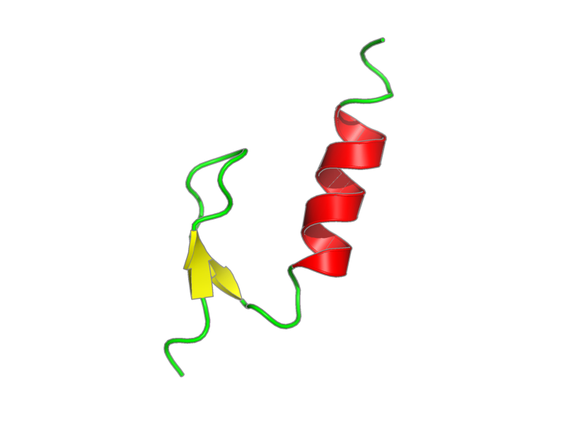
PDB ID:
Entry in NMR Restraints Grid
Validation report in NRG-CING
Chem Shift validation: AVS_full
BMRB Entry DOI: doi:10.13018/BMR15160
MolProbity Validation Chart
NMR-STAR file interactive viewer.
NMR-STAR v3 text file.
XML gzip file.
RDF gzip file.
All files associated with the entry
Citation: Bomar, Martha; Pai, Ming-Tao; Tzeng, Shiou-Ru; Li, Shawn Shun-Cheng; Zhou, Pei. "Structure of the ubiquitin-binding zinc finger domain of human DNA Y-polymerase eta" EMBO Rep. 8, 247-251 (2007).
PubMed: 17304240
Assembly members:
UBZ_domain, polymer, 39 residues, Formula weight is not available
ZN, non-polymer, 65.409 Da.
Natural source: Common Name: not available Taxonomy ID: not available Superkingdom: not available Kingdom: not available Genus/species: not available not available
Experimental source: Production method: recombinant technology Host organism: Escherichia coli Vector: pET15b
Entity Sequences (FASTA):
UBZ_domain: GSHMAAEDQVPCEKCGSLVP
VWDMPEHMDYHFALELQKS
- assigned_chemical_shifts
| Data type | Count |
| 13C chemical shifts | 126 |
| 15N chemical shifts | 36 |
| 1H chemical shifts | 258 |
Additional metadata:
Assembly:
| Entity Assembly ID | Entity Name | Entity ID |
|---|---|---|
| 1 | UBZ domain | 1 |
| 2 | ZINC ION | 2 |
Entities:
Entity 1, UBZ domain 39 residues - Formula weight is not available
corresponding to GSHM due to coloning stategy plus residues 628-662 of human polymerase eta
| 1 | GLY | SER | HIS | MET | ALA | ALA | GLU | ASP | GLN | VAL | ||||
| 2 | PRO | CYS | GLU | LYS | CYS | GLY | SER | LEU | VAL | PRO | ||||
| 3 | VAL | TRP | ASP | MET | PRO | GLU | HIS | MET | ASP | TYR | ||||
| 4 | HIS | PHE | ALA | LEU | GLU | LEU | GLN | LYS | SER |
Entity 2, ZINC ION - Zn - 65.409 Da.
| 1 | ZN |
Samples:
sample_1: UBZ domain, [U-100% 15N], 2 mM; phosphate 25 mM; KCL 100 mM
sample_2: UBZ domain, [U-100% 13C; U-100% 15N], 2 mM; phosphate 25 mM; KCL 100 mM
sample_3: UBZ domain, [U-10% 13C; U-100% 15N], 2 mM; phosphate 25 mM; KCL 100 mM
sample_conditions_1: ionic strength: 100 mM; pH: 7; pressure: 1 atm; temperature: 298 K
Experiments:
| Name | Sample | Sample state | Sample conditions |
|---|---|---|---|
| 2D 1H-15N HSQC | sample_1 | isotropic | sample_conditions_1 |
| 2D 1H-13C HSQC | sample_3 | isotropic | sample_conditions_1 |
| 2D 1H-1H NOESY | sample_1 | isotropic | sample_conditions_1 |
| 3D HCCH-TOCSY | sample_2 | isotropic | sample_conditions_1 |
| 3D HNCO | sample_2 | isotropic | sample_conditions_1 |
| 3D HNCACB | sample_2 | isotropic | sample_conditions_1 |
| 3D HNCA | sample_2 | isotropic | sample_conditions_1 |
| 3D HN(CO)CA | sample_2 | isotropic | sample_conditions_1 |
| 2D 1H-13C HSQC | sample_3 | isotropic | sample_conditions_1 |
| 3D HN(COCA)CB | sample_2 | isotropic | sample_conditions_1 |
| 3D 1H-15N NOESY | sample_1 | isotropic | sample_conditions_1 |
| 3D 1H-13C NOESY | sample_2 | isotropic | sample_conditions_1 |
Software:
XEASY, Bartels et al. - chemical shift assignment, peak picking
TALOS, Cornilescu, Delaglio and Bax - geometry optimization
CYANA, Guntert, Mumenthaler and Wuthrich - structure solution
X-PLOR NIH, Schwieters, Kuszewski, Tjandra and Clore - refinement
NMRPipe, Delaglio, Grzesiek, Vuister, Zhu, Pfeifer and Bax - processing
NMR spectrometers:
- Varian INOVA 800 MHz
- Varian INOVA 600 MHz
Download HSQC peak lists in one of the following formats:
CSV: Backbone
or all simulated peaks
SPARKY: Backbone
or all simulated peaks